


Among the many diseases that threaten human health, acute ischemic stroke (commonly known as cerebral infarction) is a major condition that severely impacts people's quality of life due to its high morbidity, disability, and mortality rates. The emergence of neurointerventional thrombectomy stents has brought new hope and breakthroughs to the treatment of this disease.
Acute ischemic stroke primarily results from cerebral vascular obstruction by a blood clot, leading to localized brain tissue ischemia and hypoxia, which in turn triggers a series of severe neurological dysfunctions. In the past, treatment options for this condition were relatively limited. While thrombolytic drugs were a common approach, their effectiveness was often unsatisfactory for strokes involving large vessel occlusion. The advent of neurointerventional thrombectomy stents has changed this situation.
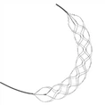
Structurally, neurointerventional thrombectomy stents are typically made of specialized metal materials or polymers, offering excellent flexibility and support. Designed like a "small net bag," they deploy smoothly within the blood vessel and adhere closely to the clot. During the actual procedure, the surgeon first uses techniques such as angiography to precisely determine the location and size of the clot. The surgeon then performs a puncture in the patient's groin and inserts a thin guide catheter into the femoral artery, establishing a safe passage for subsequent procedures.
Next, under the guidance of the guidewire, the microcatheter loaded with the stent retriever is carefully delivered to the cerebral vascular area where the blood vessel is located. This step requires the doctor to have superb skills and extensive experience, because cerebral blood vessels are very delicate and complex in structure, and the slightest carelessness can cause damage to the blood vessels. When the microcatheter reaches the thrombus, the doctor will release the stent retriever and allow it to expand within the thrombus. The mesh design of the stent retriever can fit well with the thrombus. After waiting for a while to ensure that the stent and the thrombus are fully integrated, the doctor will slowly pull the stent retriever out of the body along with the thrombus, thereby restoring the blocked blood vessel and restoring blood supply to the brain.
Neurointerventional thrombectomy stent therapy has many significant advantages over traditional treatment methods. First, it can directly remove the thrombus from the blood vessel, and has a higher recanalization rate for large vessel occlusions. Studies have shown that the use of thrombectomy stents can restore blood flow to the brain in a short period of time, greatly reducing the number of nerve cells that die due to ischemia, thereby reducing the risk of severe disability in patients. Secondly, this treatment method is a minimally invasive surgery and causes relatively little trauma to the patient. Compared with traditional craniotomy, it does not require opening the skull, and the treatment operation can be completed through a small puncture point, and the patient's postoperative recovery time is also significantly shortened.
Of course, no medical technology is perfect. Neurointerventional thrombectomy with stents also has certain risks and limitations. For example, during the surgical procedure, blood vessels may be damaged, causing rupture and bleeding; during the thrombus removal process, it may also fall off and enter other blood vessels with the blood flow, causing new blockages. In addition, thrombectomy with stents has a strict time window limit and is generally recommended to be performed within 6-24 hours after onset. The earlier the treatment, the better the effect. Therefore, brain tissue is very sensitive to ischemia. Every minute and every second of delay may lead to more nerve cell death, affecting the patient's prognosis.