

Intracranial aneurysm is a cerebrovascular disease that seriously threatens human health. Rupture can lead to serious consequences such as subarachnoid hemorrhage, with extremely high disability and mortality rates. Coil embolization, as one of the important minimally invasive methods for the treatment of intracranial aneurysms, has been widely used in clinical practice due to its advantages of less trauma and faster recovery.
Before surgery, doctors need to fully assess the patient's physical condition. Ask the patient's medical history in detail, including whether the patient has underlying diseases such as hypertension, diabetes, heart disease, and whether there is a tendency to bleed. Perform a neurological examination to understand the patient's state of consciousness, neurological deficits, etc. Through imaging examinations such as cranial CT, MRI, and cerebral angiography (DSA), the location, size, shape, neck width, and other key information of the aneurysm can be accurately determined, which is crucial for formulating surgical plans and selecting appropriate coils.
At the same time, prepare appropriate interventional instruments, such as vascular sheaths, guide catheters, microcatheters, microguidewires, etc. According to the characteristics of the aneurysm, choose coils of different specifications, including the diameter, length, and number of coils of the coils, etc. At the same time, prepare the drugs that may be used during the operation.
The surgical procedure includes vascular puncture and access establishment, microcatheter placement, coil embolization and postoperative treatment.
First, vascular puncture and access are established. The patient is placed in a supine position, and routine disinfection and draping are performed. Under local anesthesia, the right femoral artery is usually selected for puncture. The puncture needle is inserted into the femoral artery using the Seldinger technique. After successful puncture, a guide wire is introduced, and the vascular sheath is placed into the femoral artery along the guide wire. The vascular sheath provides a safe channel for subsequent catheter operations. Through the vascular sheath, the guide catheter is slowly delivered to the appropriate position of the affected internal carotid artery or vertebral artery under the guidance of the guide wire as a support for subsequent microcatheter operations.
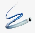
Secondly, under the real-time monitoring of DSA, the micro-guidewire is combined with the micro-catheter, and the micro-catheter is carefully passed through the guide catheter and gradually inserted into the aneurysm cavity. This process requires the operator to have fine operation skills and closely observe the position and direction of the micro-catheter to avoid damaging the blood vessel wall. After the micro-catheter is in place, a small amount of contrast agent is injected to confirm that the tip of the micro-catheter is in the aneurysm cavity and there is no abnormality such as contrast agent spillage.
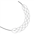
Then, according to the size and shape of the aneurysm, select the appropriate coil to start embolization. The coil is slowly pushed into the aneurysm cavity through the microcatheter, so that it can coil into a tight ball-like structure in the aneurysm cavity. During the release of the coil, DSA monitoring is continuously performed to ensure the good distribution of the coil in the aneurysm cavity and to prevent the coil from falling out and entering the tumor-bearing artery. After each coil is released, angiography is performed to evaluate the packing effect of the coil and the blood flow in the aneurysm cavity. According to the angiography results, select subsequent coils of appropriate specifications to continue packing until the aneurysm cavity is completely or nearly completely occluded, while ensuring the patency of the tumor-bearing artery.
After the coil embolization is completed, the microcatheter and guide catheter are withdrawn, and the puncture site is compressed to stop bleeding. Generally, after 15-30 minutes of compression, confirm that there is no bleeding, and use a vascular closure device or pressure bandage to stop bleeding. The patient needs to lie flat for 12-24 hours after the operation, and closely observe whether there is bleeding, hematoma formation, and dorsalis pedis artery pulsation at the puncture site. At the same time, monitor the patient's vital signs, neurological symptoms and changes in signs. Give the patient appropriate anticoagulant and antiplatelet therapy to prevent thrombosis, but pay attention to monitoring coagulation function to avoid bleeding complications. For possible complications such as cerebral vasospasm, give appropriate drug treatment.
Intracranial aneurysm coil embolization is a surgery with high technical requirements and delicate operation. Strictly following the standardized surgical process, from comprehensive preoperative evaluation and preparation, to precise intraoperative operation, to careful postoperative care and monitoring, every link is important. Through the rational use of this technology, aneurysms can be effectively occluded, the risk of aneurysm rupture and bleeding can be reduced, the patient's prognosis can be improved, and better treatment effects can be brought to patients with intracranial aneurysms.