

Endovascular treatment of intracranial aneurysms has been growing in popularity since the first successful coil embolization procedure in 1991. This minimally invasive technique involves the placement of small coils inside the aneurysm sac to prevent further growth and reduce the risk of rupture. Compared to surgical clipping, coil embolization offers a shorter hospitalization, quicker recovery time and fewer complications. Clinicians interested in mastering this technique should have a good understanding of the basic steps involved in aneurysm coiling.
Patient Selection
The first step in aneurysm coiling is patient selection. Not all aneurysms are suitable for this technique. Patients with symptomatic or ruptured aneurysms are typically treated with surgical clipping instead of coil embolization, as the latter may not be able to immediately stop the bleeding. Additionally, certain anatomical characteristics such as large size or wide neck may make aneurysm coiling technically challenging or impossible. A thorough evaluation of the patient's clinical history, imaging studies, and medical condition should be performed to determine whether they are a good candidate for this procedure.
Access Site Selection
Once the patient has been deemed a suitable candidate, the next step is access site selection. The most common access site is the femoral artery in the groin. This site is chosen because it provides a relatively straight path to the brain and is less likely to cause complications such as bleeding or hematoma. However, when the aneurysm is located in the posterior circulation, access through the radial artery in the wrist or the brachial artery in the arm may be preferred.
Catheterization and Aneurysm Selection
After access site selection, a specialized catheter is guided through the arterial system towards the aneurysm site. Using fluoroscopic guidance, the catheter is navigated to the cerebral artery that supplies blood to the aneurysm. Various imaging techniques such as digital subtraction angiography (DSA) or three-dimensional rotational angiography (3DRA) may be used to obtain a better visualization of the aneurysm and surrounding vasculature. Once the aneurysm has been identified, the size, shape, and location are assessed, and an appropriate coil is selected.
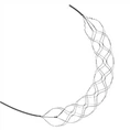
Coil Embolization
The coil is advanced through the catheter and into the aneurysm sac. The coil is then released and expands to fill the aneurysm space. Multiple coils may be used to pack the aneurysm cavity as densely as possible, thereby minimizing the chance of blood flow into the aneurysm sac. Once embolization is complete, a follow-up angiogram is performed to verify the placement of the coils and determine the degree of aneurysm occlusion.
Post-Procedure Care
Following the procedure, careful monitoring is required to ensure that complications such as bleeding, thrombosis, or vasospasm do not occur. Patients are typically observed in the hospital for 24 to 48 hours to ensure that there are no immediate complications. After discharge, patients are advised to avoid strenuous activities and to consult a physician if they experience any neurological symptoms such as headache, numbness, or weakness.
Conclusion
Aneurysm coiling is a safe and effective treatment option for patients with certain types of intracranial aneurysms. This minimally invasive procedure offers numerous advantages over traditional surgical clipping, such as a shorter hospital stay and faster recovery time. However, this procedure requires careful patient selection, access site selection, and skilled catheterization and embolization techniques. With the proper training and equipment, endovascular specialists can achieve excellent outcomes for their patients.