

Selection of the appropriate sheath and catheter, and the proper use of relevant techniques in a certain sequence, are all critical to the success of any neurovascular intervention and are key to avoiding catastrophic complications. The choice of device depends on the anatomical path to the blood vessels in the target area and the type of interventional plan.
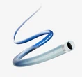
The sheath is a catheter composed of a one-way valve and an injection end. It is commonly used for blood vessel puncture of the femoral artery, radial artery and brachial artery. The sheath allows rapid exchange of catheters and equipment with little potential damage to the vascular access site. In a randomized controlled trial, the use of an arterial sheath reduced the incidence of bleeding at the femoral artery puncture site during operation and improved the convenience of catheter operation without increasing the incidence of complications on the puncture side. Short sheaths (10 to 13cm) are often used. And it’s available diameters ranges from 4 to 10F. During neuroangiographic procedures, the sheath needs to be continuously pressurized with heparinized saline at arterial pressure. A long sheath (25 cm) may be selected when atherosclerosis or tortuosity of the iliofemoral artery precludes catheter delivery. An 80cm or 90cm long sheath can reach the carotid artery or subclavian artery and be used as a stabilizing device to support the guiding catheter or for large-lumen guiding catheters.
Catheters used for neurovascular intervention are divided into diagnostic catheters and guiding catheters. These catheters can reach the target blood vessels on the aortic arch and allow microcatheters to reach the intracranial circulation. Hydrophilic guidewires or microguidewires are used to help these catheters reach the target site.
Diagnostic catheter: The standard catheter used for cerebral angiography is a 4F or 5F tapered angle catheter. The usual length of the catheter is 90cm to ensure sufficient length outside the sheath. 4F or 5F catheters can be used in patients with bovine aortic arch tortuosity. A 5F catheter can also be used to access the right subclavian artery or right vertebral artery. The diagnostic catheter is often advanced under the support of a hydrophilic guidewire. The path of the guidewire tip should be tracked under direct fluoroscopy from the beginning of femoral artery puncture. The guidewire should always be 8 to 10cm longer than the catheter to avoid vessel wall dissection. . Path planning techniques should be used when accessing the vertebral, internal, and external carotid arteries.
Guiding catheter: The guiding catheter provides a stable platform through which the microcatheter can reach distal small vessels during interventional therapy. The 5F guiding catheter allows placement of a microcatheter with sufficient clearance for irrigation and contrast injection. 6F or 7F guiding catheters are used for patients who require greater support. Some catheters are non-hydrophilic, are more stable within the vessel, provide a good platform in tortuous vessels, and have a larger lumen. The balloon of the balloon guiding catheter can block the proximal blood flow and prevent embolism in the distal blood vessels, especially during carotid artery interventional treatment. The lumen of these catheters is relatively small, only 80cm in length. The catheter has a soft, atraumatic tip, but it is hydrophilic and slides easily. A sheath or guiding catheter that provides stiff, stable support.
Details of the use of guiding catheters play a key role in the success of intracranial embolization treatment because they provide a stable platform for soft and flexible microcatheters to enter intracranial blood vessels. The catheter can be inserted directly into the target vessel in young patients without tortuosity and arteriosclerosis. In patients with tortuous anatomy, arteriosclerosis, or myofibrillar dysplasia, an exchange guidewire should be used for exchange. The guiding catheter should be guided into the carotid and vertebral arteries using the path map. The further apart it is placed, the more stability it provides. In the carotid artery system without tortuosity and disease, it is recommended to place the head end of the guiding catheter in the vertical segment of the petrous part of the internal carotid artery. In the obviously tortuous neck of the internal carotid artery, the tip of the guiding catheter only needs to be placed just above the proximal end of the curve. The ideal location for the tip of the vertebral artery guiding catheter is distal to the extracranial segment of the vertebral artery, usually at the first bend. When the guiding catheter is in place, contrast agent is injected through the guiding catheter (under fluoroscopy) to check the morphology of the blood vessels around the catheter tip and check for vasospasm or vascular dissection around the catheter tip. If vasospasm and flow restriction due to the catheter tip occur, withdrawing the catheter 1 mm is often sufficient to restore flow. Continuous lavage of the guiding catheter with heparinized saline is important to avoid thrombosis and distal embolization. It is also important to monitor the position of the guiding catheter under regular fluoroscopy during microcatheter entry and interventional procedures to ensure that the guiding catheter is in the appropriate position.
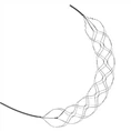
Microcatheters can reach the intracranial circulation coaxially through the guiding catheter. They are divided into guidewire-guided microcatheters, blood flow-guided microcatheters, or controllable guidewire-guided microcatheters. Guidewire-guided microcatheters are most commonly used. These microcatheters vary in length, internal and external diameter, and shape. The Tranvi Microcatheter is compatible with dimethyl sulfoxide (DMSO, required for liquid embolic agents). Microcatheter selection depends on the following: the type of device and embolic agent delivered through the microcatheter, the diameter relative to the inner diameter of the guiding catheter that will allow injection through the guiding catheter, and the anatomy or tortuosity that must be overcome to reach the target site. A two-point labeled microcatheter is required to use a releasable coil, rather than a single-marked microcatheter. These two marks make the distal 3cm of the microcatheter slightly harder than the corresponding part of the single-marked microcatheter.
Subtle differences in the use of guidewire-guided microcatheters: Bidirectional path maps are crucial for precise superselection of microcatheters and monitoring the position of microcatheters during operation. During the operation, heparinized saline must be used to continuously flush the guiding catheter and microcatheter. All guidewire-guided microcatheters have a hydrophilic coating, are packaged in a plastic hoop, and can be flushed with sterile heparinized saline to hydrate the coating. Connect the microcatheter to the rotary hemostasis valve and remove the air from the microcatheter with heparinized saline. Use a guidewire guide to insert the microguidewire into the rotary hemostasis valve. The twist controller is fixed on the proximal end of the microguidewire, and the guidewire is controlled by rotating the curved head end of the distal end of the guidewire. The tip of the microcatheter can exceed the microguidewire in straighter blood vessel segments, thereby reducing vessel damage or perforation. At sharp bends or branches of blood vessels, the microguidewire should be rotated and passed carefully. When the microcatheter reaches the desired location, gently pull and withdraw the microguidewire. Observe the tip of the microcatheter under fluoroscopy and withdraw the microguidewire, because removing the microguidewire will release the energy accumulated on the microcatheter, allowing the microcatheter to advance forward. Injecting a small amount of contrast agent through the microcatheter can determine the position and patency of the microcatheter. It is necessary to pay attention to the rotating hemostatic valve connected to the microcatheter (and guiding catheter) throughout the process to determine whether there are thrombus or air bubbles.
6. Risk prevention: Detailed assessment of the patient's preoperative and intraoperative anatomy, the goals of interventional treatment, and the mastery of the characteristics and performance of various sheaths and catheters are very important to the success of neurovascular endovascular operations and are also the key to avoiding complications.